
CAROTID BODY TUMOURS (includes operative photographs)
WARNING: this article includes graphic medical photography of a Carotid Body Tumour surgery.
Carotid body tumours are rare and usually present as a non-painful swelling in the upper neck.
They grow from specialised nerve cells found in the junction between the two main arteries in the neck, the internal and external carotid arteries. Due to this anatomical position carotid body tumours are close to, and can grow around, both these blood vessels and also important nerves that help the tongue, throat and voice box to function.
A comprehensive history, head and neck examination and appropriate investigations are important to ensure the correct diagnosis is made. Please feel free to contact Dr Farrell’s rooms if you have any enquiries concerning lumps in the neck.
The recommended treatment of carotid body tumours is surgical removal.
A highly specialised surgical unit which includes an experienced head and neck surgeon, vascular surgeon and interventional radiologist are essential to ensure a safe and complete removal.
Dr Farrell, who has experience with this surgery, recently removed a carotid body tumour, preserving the two main arteries and maintaining the function of the neighbouring important nerves (please see pictures below). After surgery the patient is able to talk, swallow and socialise normally.
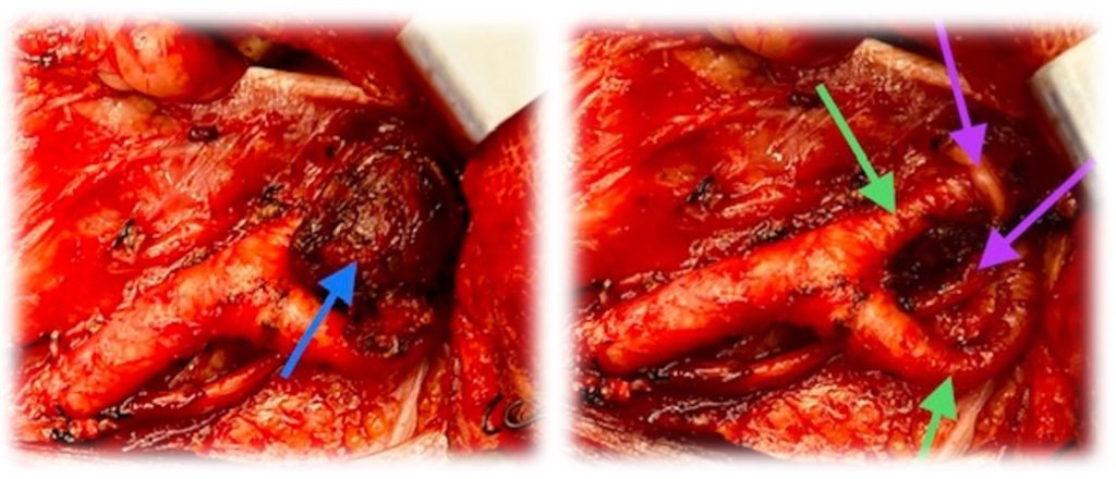
The picture on the right shows the tumour removed and the main arteries, green arrows, and large nerves, purple arrows, lying unharmed and intact underneath.
Please contact Dr Farrell’s rooms for further details.
LATEST POST
- MULTIPLE TRANS ORAL ROBOTIC SURGERIES Completed with Excellent Functional and Oncological Results
- CAROTID BODY TUMOURS (includes operative photographs)
- LARYNGEAL COBLATION – A NEW SURGICAL TECHNIQUE
- LOW IRON LEVELS FOUND IN CHILDREN REQUIRING TONSIL AND ADENOID SURGERY
- IMMUNITY & SKIN CANCERS OF THE FACE, SCALP & NECK
- NEW INNOVATIONS IN THE TREATMENT OF SNORING
- MAJOR HEAD & NECK SURGERIES ARE INCREASING
- TRANS ORAL ROBOTIC SURGERY
- VIDEO ASSISTED NASENDOSCOPE
- REVOLUTIONARY NEW ANAESTHETIC TECHNIQUE